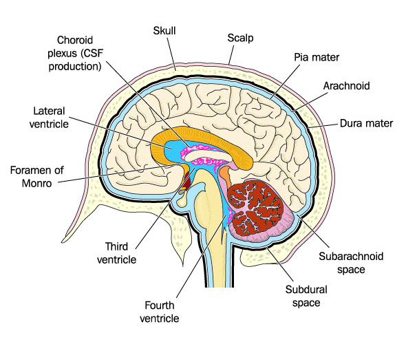
Choroid plexus diagram Geeky Medics

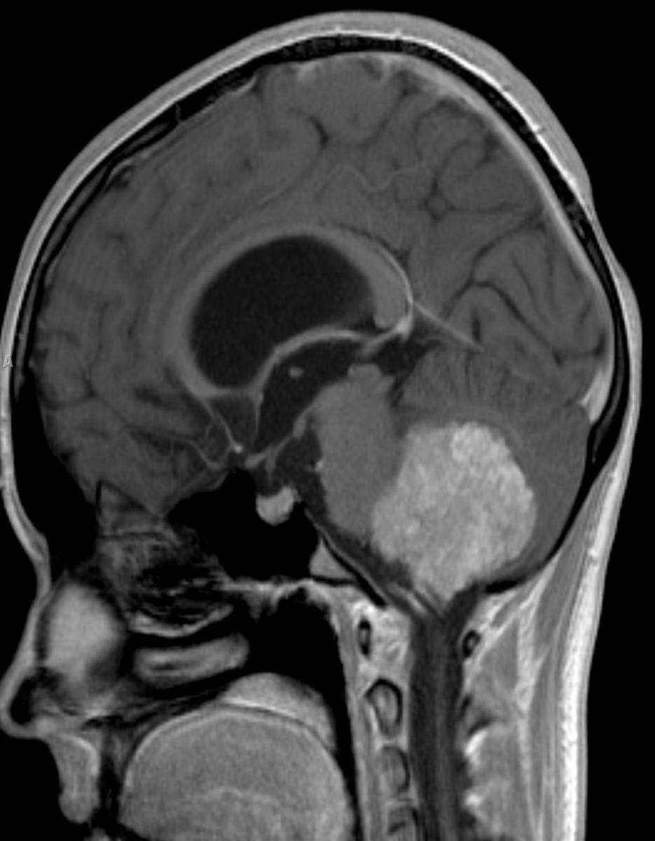
Choroid Plexus Xanthogranuloma Neuro MR Case Studies CTisus CT Scanning
choroid plexus / intraventricular metastases adjacent parenchymal tumors (e.g. glioblastoma) Secondary Secondary causes of intraventricular hemorrhage include: extension from other intracerebral hemorrhages hypertensive hemorrhage, especially basal ganglia hemorrhage (common)

Xanthogranulomas of the choroid plexus Image
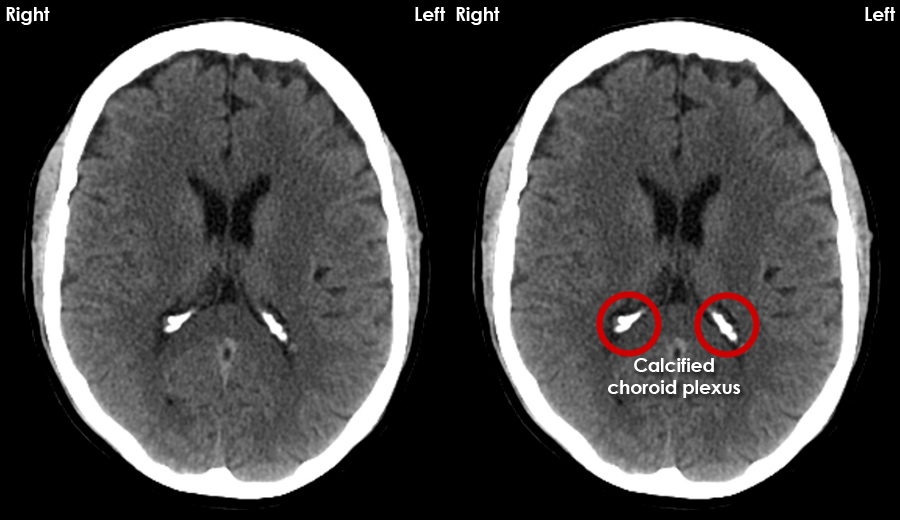
Physiologic calcification of the choroid plexus increases in frequency and extent with age. As demonstrated in this report, it is visualized nine to 15 times more frequently with computed tomography (CT) than with plain skull radiography. Calcification involving the temporal horns is associated with neurofibromatosis. Young patients with exuberant calcification in the region of the glomerula.
Pictures Of Choroid Plexus
The choroid plexus lies at the interface between the cerebrospinal fluid and the systemic circulation, its capillaries have a fenestrated epithelium that serves as a way through which infectious organisms may gain access to the CNS 1,2 . Radiographic features CT and MRI
CT Brain Anatomy Calcified structures
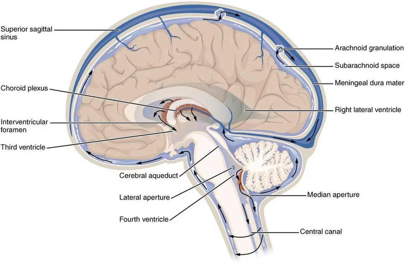
The choroid plexus, or plica choroidea, is a plexus of cells that arises from the tela choroidea in each of the ventricles of the brain. [1] Regions of the choroid plexus produce and secrete most of the cerebrospinal fluid (CSF) of the central nervous system.
Onset of Cognitive Dysfunction in Systemic Lupus Erythematosus and Selective Involvement of the
Choroid plexus cysts are of concern if the cysts are large (>1 cm) (controversial evidence), bilateral, multiple, and associated with structural abnormalities when the maternal age is ≥32 years, or if the maternal serum screening results are abnormal.
Choroid plexus diagram Geeky Medics
The choroid plexus (CP) can be seen anywhere throughout the ventricular system with the exception of the cerebral aqueduct. It consists of numerous capillary-rich villi (with a blood flow of nearly ten times that of the cerebral cortex), which in turn are composed of a type of ependymal cells. The purpose of the choroid is to serve as a sort of.
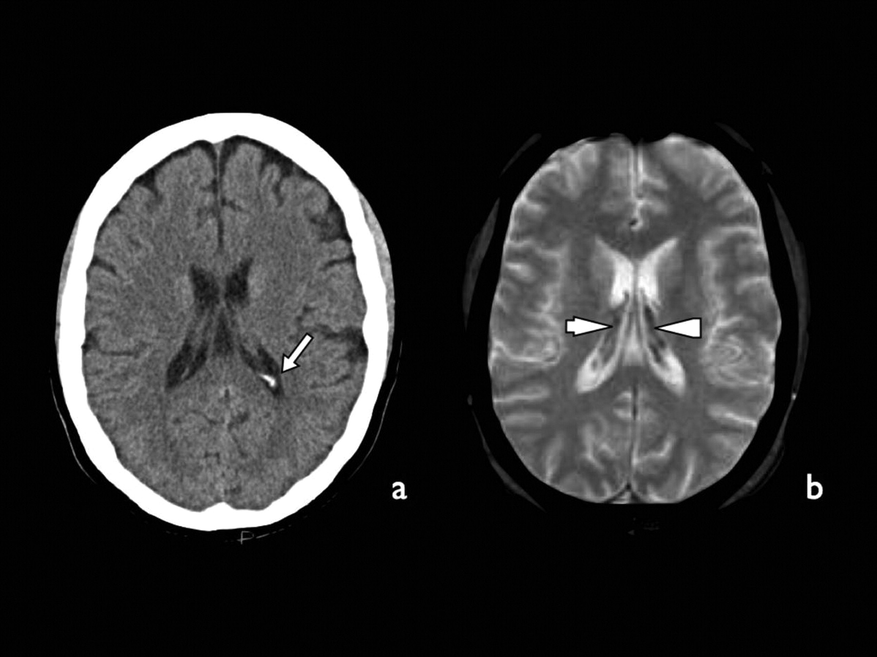
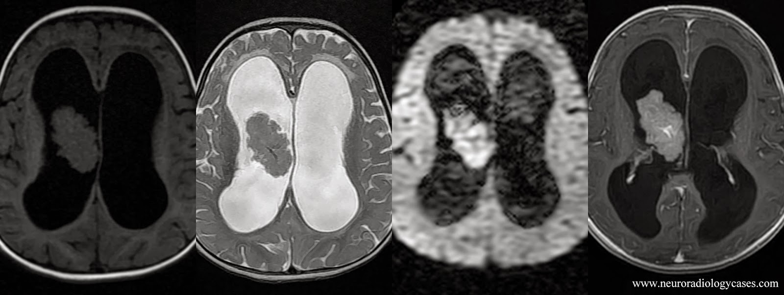
Neuroradiology Cases Choroid plexus cyst MRI
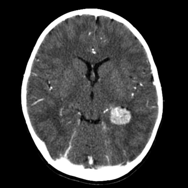
Choroid plexus cysts are most frequently located in the trigones of the lateral ventricles, but can occur throughout the ventricular system. They are typically less than 1 cm in diameter and are round or ovoid in shape. On CT, choroid plexus cysts usually demonstrate CSF density but can occasionally appear hyperdense relative to the CSF.
Dr Balaji Anvekar FRCR Choroid Plexus Papilloma MRI
Choroid plexus carcinoma is a rare type of brain cancer that happens mainly in children. Choroid plexus carcinoma begins as a growth of cells in the part of the brain called the choroid plexus. Cells in the choroid plexus produce the fluid that surrounds and protects the brain and spinal cord.

Choroid Plexus Papilloma Neuro MR Case Studies CTisus CT Scanning
The choroid plexus is a complex network of capillaries lined by specialized cells and has various functions. One of the primary functions is to produce cerebrospinal fluid (CSF) via the ependymal cells that line the ventricles of the brain.
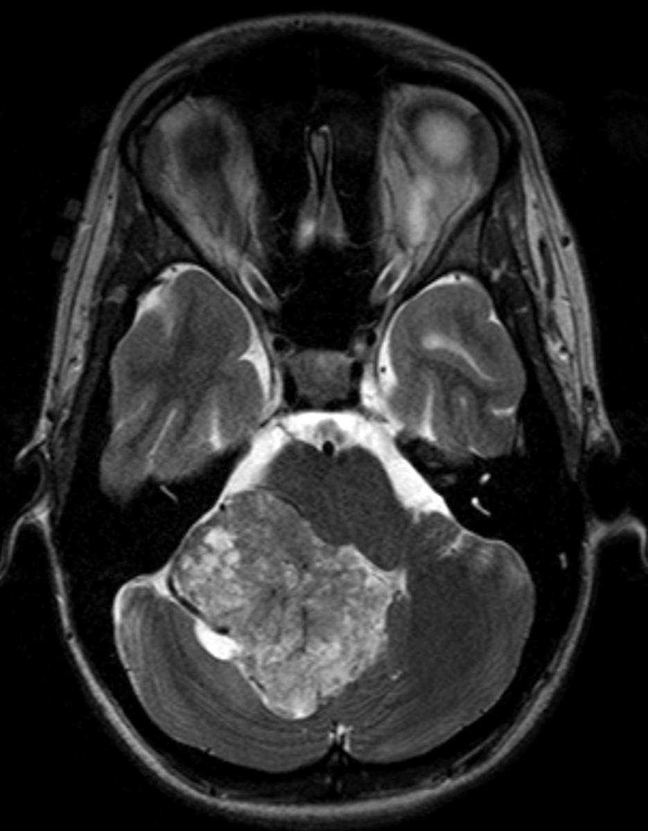
Choroid Plexus Papilloma Neuro MR Case Studies CTisus CT Scanning
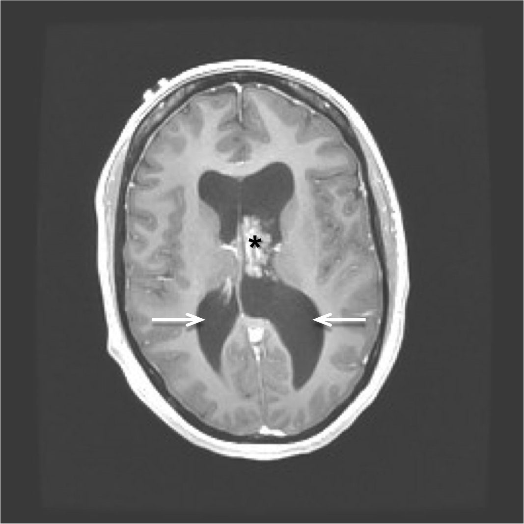
Choroid plexus xanthogranulomata are common, incidental and almost invariably asymptomatic lesions. It is unclear in much of the literature whether they represent a distinct entity from adult choroid plexus cysts, but they share imaging characteristics and are only likely to be distinguishable on autopsy.
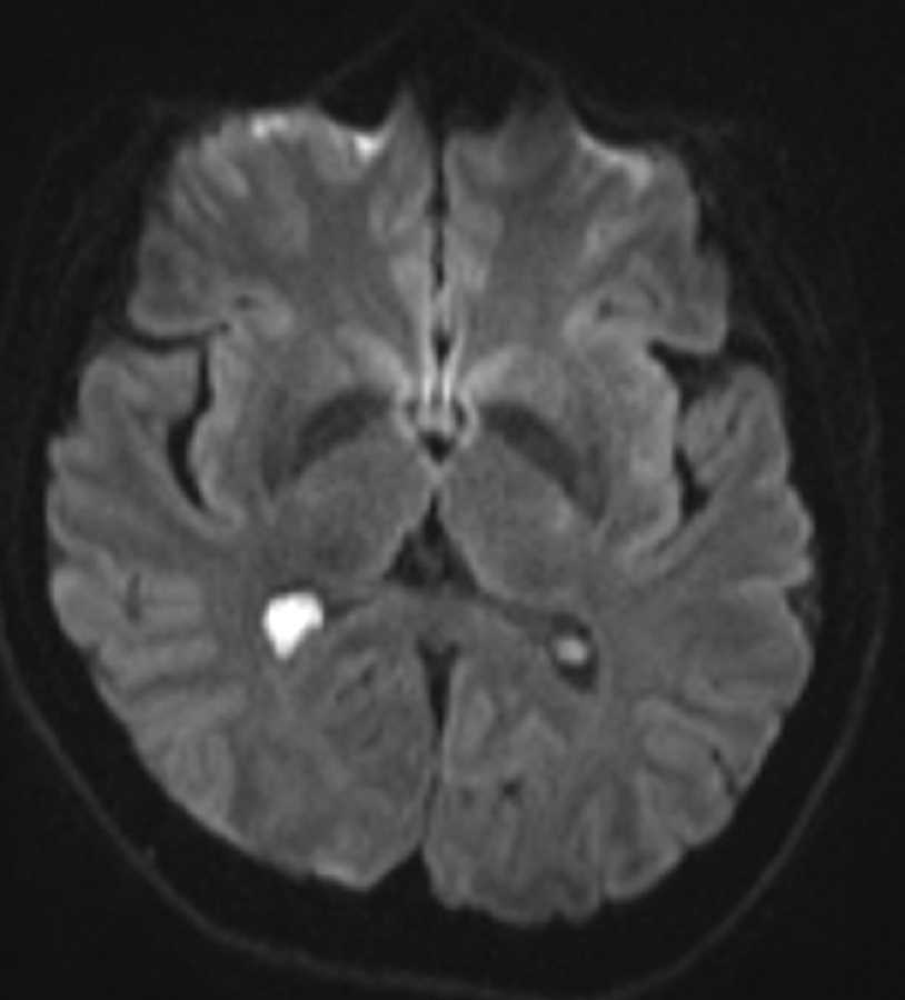
Choroid Plexus Xanthogranuloma Neuro MR Case Studies CTisus CT Scanning
Choroid plexus tumors are grouped in three grades based on their characteristics (grade 1, 2, or 3, also written as grade I, II, or III) . Grade 1 choroid plexus papilloma are low grade tumors. This means the tumor cells grow slowly. Grade 2 atypical choroid plexus papilloma are mid-grade tumors. This means the tumors have a higher chance of.
Choroid plexus MedCBL Inc.
The choroid plexus is a complex tissue configuration made up of epithelial cells, capillaries (tiny blood vessels), and connective tissue that lines the ventricles of the brain. Its function first and foremost is to secrete cerebrospinal fluid (CSF), a clear fluid that protects the brain and spinal cord. It has other important functions as well.
Choroid Plexus Papilloma Neuro MR Case Studies CTisus CT Scanning
The choroid plexus is located within the cerebral ventricles and is made of choroidal epithelial cells (type of ependymal cell), loose connective tissue ( tela choroidea ), and permeable capillaries. It produces cerebrospinal fluid . The choroid plexuses also form the blood-CSF barrier alongside arachnoid and arachnoid villi 2. Gross anatomy
Choroid plexus papilloma wikidoc
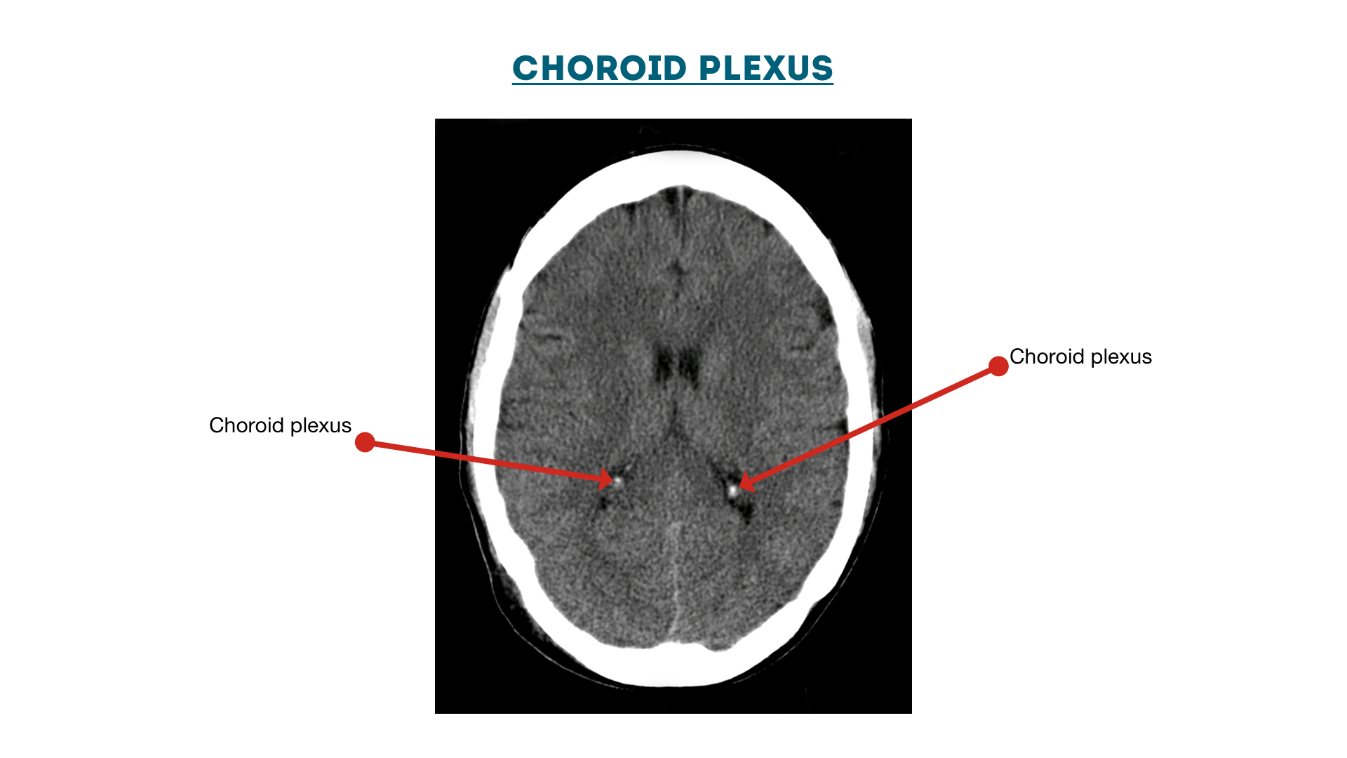
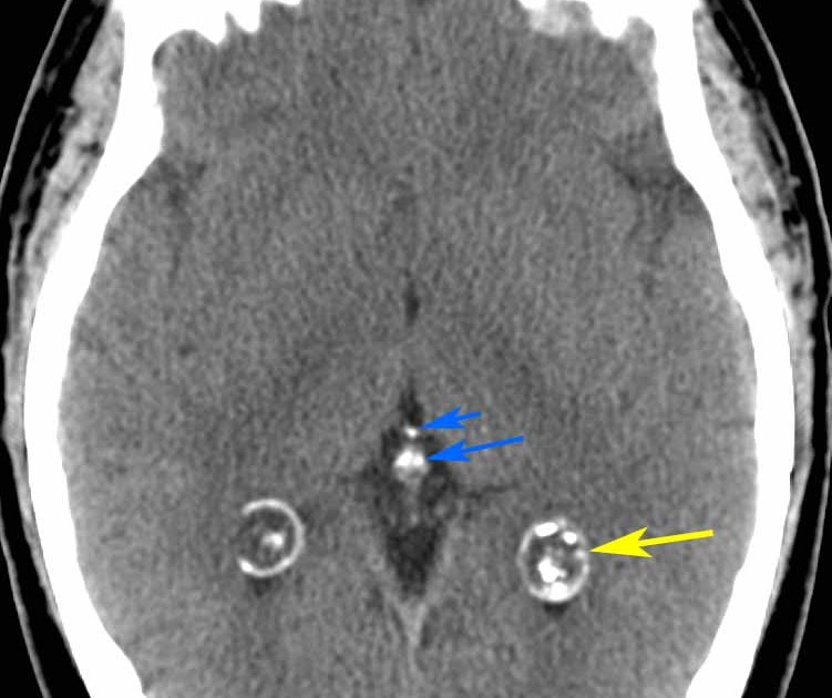
choroid plexus a very common finding, usually in the atrial portions of the lateral ventricles (choroid glomus) 1 calcification in the third or fourth ventricle or in patients less than nine years of age is uncommon basal ganglia most commonly in the globi pallidi
RiT radiology Calcified Choroid Plexus Cysts
CT Brain Anatomy Calcified structures Key points Commonly calcified structures of the brain include the choroid plexus, pineal gland, basal ganglia and falx Use of CT 'bone windows' is helpful in differentiating calcified structures from acute haemorrhage There are several structures in the brain which are considered normal if calcified.
Choroid Plexus The Definitive Guide Biology Dictionary
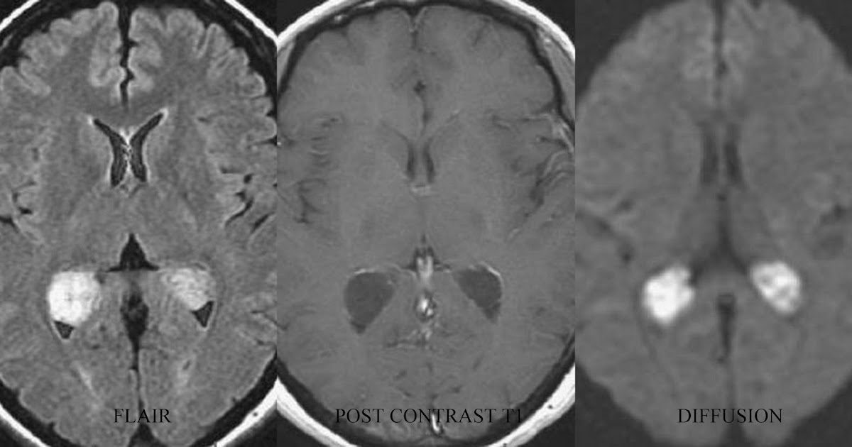
Summary: Infarction of the choroid plexus may result from ischemia in the distribution of the medial posterior choroidal artery. Diffusion-weighted imaging may depict this unusual stroke syndrome. The clinical and radiologic aspects of this rare condition are discussed taking into consideration the anatomy and pathophysiology of the choroid plexus.
